
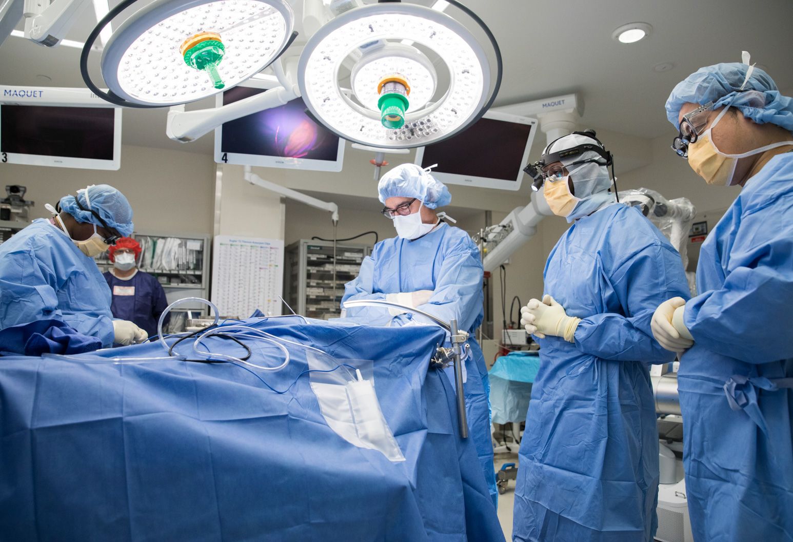
Depending on how fast the tumor grows and which area is affected, the preparation can take several weeks and neurosurgery medical instruments are being used for it.
Stereotaxic And Neuro-Navigation Before Surgery
If the tumor grows in a very unfavorable location, is very large, and is removed in several sessions, or if the patient is in a poor general condition, the surgeon will remove part of the tumor with the help of stereotaxic or neuro-navigation. In the context of stereotaxic, the tumor is calculated and targeted using mathematical calculations. Neuro-navigation calculates the position of the tumor using magnetic fields and ultrasonic pulses. A computer stores the data and creates a three-dimensional image. The surgeon uses this image as a guide during the operation and guides the instruments through real-time CT and MRI images. This makes it possible, on the one hand, to use the computer to make various brain structures visible and to protect them.
On the other hand, the surgical instruments and the position of the tumor can be displayed on a screen at the same time so that the surgeon can take a sample or remove part of the tumor.
Neuro-Mapping On The Conscious Mind Before Surgery
The operation, known in technical jargon as awake craniotomy, is mainly carried out on people whose tumor is in motor or linguistic areas. A wake-up operation is also performed as part of epilepsy and Parkinson’s therapy. This requires a so-called neuro-mapping beforehand. A map of the brain is created in a magnetic resonance tomograph. During these recordings, the patients have to carry out various tests, for example, counting, arithmetic, reading, reciting days of the week, solving memory tasks, etc. The magnetic resonance tomograph shows which areas in the brain are activated. This allows the surgeon to better delimit the healthy areas from the tumor in a later operation.
If it is tumor tissue, it no longer fulfills a function, and the surgeon can better assess the limit and reduce the risk of surgery. After hemostasis and an imaging control, the three meninges and the skull are closed. The patient is transferred to a neurosurgical monitoring station, where he wakes up after the anesthesia.